By James Martin, with files from Alison Ramsey
Structure is central to all science: proteins and biology, molecules and chemistry, the crystal lattices and physics. But what these structures look like while undergoing crucial transformations—say, during protein function or chemical reactions—is still the stuff of imagination. “We all make mental pictures of what’s happening,” says Bradley Siwick, “but we don’t know.” Siwick wants to pull back the curtain on this mystery and take pictures of these blazingly fast changes. His femtosecond laser and electron microscopy laboratory, three years in the making, just might do the trick.

Familiarity doesn’t preclude mystery.
Humans and horses, for example, go way back. At least 16,000 years, according to cave paintings. We care for horses, ride them, rely on them, bet on them. They are a familiar sight (and smell). We know horses. So it’s odd that, up until 1878, we had no idea whether all four of a galloping horse’s hooves ever left the ground at the same time—a knowledge gap that was the stuff of serious debate. It took an Englishman named Eadweard Muybridge and a newfangled process known as photography to settle the question. (The short answer is yes, they do.)
Humans and atoms, too, go way back. Around 400 BC, Democritus argued that unbreakable particles were the building blocks of everything. It took some convincing, but by the time the 20th century rolled around, scientists had embraced the basic notion of these “atomos.” McGill professor Ernest Rutherford famously developed the nuclear model, which held that each atom was made up of an incredibly dense nucleus (itself made of protons and neutrons—themselves divisible into quarks, so Democritus was a bit off ) surrounded by one or more electrons. Rutherford even went so far as to cleave an atom in half, ushering in the atomic age and earning a Nobel Prize.
Like horses, we know atoms.
Most of the time.
Modern physics has its own horse hoof quandary: How are the atoms inside a molecule arranged during a chemical transformation? And how are the atoms in a material arranged through the transformation between phases? Using a range of well-established techniques, such as X-ray diffraction, nuclear magnetic resonance and electron microscopy, we know the equilibrium structure of many molecules and materials before such transformations, and we know the equilibrium structure of the results. It’s what happens during that change, all in a matter of 10-13 seconds, that’s the big question mark.
“What do we know about these short-lived transient states?” muses McGill researcher Bradley Siwick. He shakes his head. “Not very much.”
Now he just might be on the verge of counting the hooves.
* * *
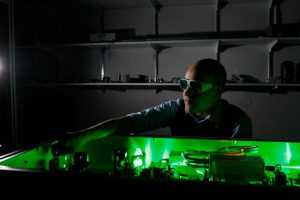
An assistant professor in the Departments of Chemistry and Physics, Siwick believes photography, in a manner of speaking, is the way to pin down a trans forming atom’s hooves. But, while Eadweard Muybridge merely had to set up a few cameras alongside a racetrack, Siwick has had to carefully erect a maze of lasers, amplifiers and lenses together with a specially designed electron microscope on a vibration-free steel table in the basement of McGill’s Otto Maass Chemistry Building. It took three years of painstaking precision to set up the rig. Now Siwick is getting down to making movies of atoms in action.
The big problem is that this action moves really, really quickly. Atoms can change position by one angstrom in mere femtoseconds. These measures, although incredibly tiny, are, in Siwick’s world, huge: An angstrom is approximately the distance between atoms in matter; a femtosecond is to one second as one second is to 60 million years. So, yes, atoms are fast. To bottle this particular lightning, one needs a camera with a correspondingly fast shutter speed. In this case, the “camera” is actually the combination of an ultrafast laser and a specially designed electron microscope.
Developed in the 1980s, the ultrafast, or femtosecond, laser produces brief pulses of light, not the continuous stream found in laser pointers and the like. The pulse is crucial because, as Siwick says, it “allows us to interact with molecules and materials ‘instantaneously.’ You can dump energy into a sample before its atoms have time to respond.” Siwick’s apparatus splits each pulse into two beams: one beam of photons that can be tuned to virtually any wavelength, from ultraviolet to infrared, and another that is transformed into a pulse of electrons (through the well-known photoelectric effect). The photons initiate a chemical reaction by exciting the molecules while the electrons pass through the molecules, producing a scattering pattern that provides an indirect, yet accurate, document of the atomic arrangement at only that instant. The result is an atomic-level view of the fleeting structural changes that make up chemical reactions. (All this occurs faster than literally anything else in the world.) Put together enough of these sequential snapshots and, in the fine tradition of “flip book” animations doodled in the margins of a textbook, you’ve got a movie. “If we can make measurements faster than atoms can move, it opens up amazing opportunities,” says Siwick. “We’ll be able to actually watch chemistry as it occurs.”
* * *
When Siwick founded his McGill lab four years ago, his team was one of just a few pioneering the field; now there are some 20 groups racing to understand molecular structure in flux. It’s a competitive world, but Bradley Siwick is a competitive guy. He was a high school hockey and football star hoping to obtain a sports scholarship in the U.S.—his dream was to study medicine—when he broke his neck while ski-jumping near his hometown of Toronto. Just four vertebrae short of total below-the-neck paralysis, he refocused his energies, becoming an internationally ranked wheelchair tennis player and studying commerce. He loved the tennis (he toured the European circuit and was part of the wheelchair equivalent of Canada’s Davis Cup team) but hated accounting. A required physics course, however, sparked his imagination. Instead of the “really boring old stuff about balls rolling down an inclined plane” that he had to suffer through in high school, modern physics fascinated him. Seven years later, his University of Toronto doctoral dissertation earned the rare distinction of being highlighted in a Science magazine cover story. His thesis documented his development of a prototype molecular movie camera and its use in studying the changing atomic structure of melting aluminum.
Setting up his McGill lab has taken time. He and his team of graduate students—Chris Godbout, Robert Chatelain and Vance Morrison—had to custom-build more than half of the equipment in the laboratory. During this period they designed a “temporal lens” for electron pulses, which will result in an overall performance increase of a factor of 10,000 compared to the current state-of-the-art. (To put this achievement in the proper perspective, consider the dictum of experimental science: “Anything better than a factor of 2 is worth fighting for.”) “Some graduate students are horrified by having to build their own experiment,” Siwick grins. “These kinds of projects draw the really ambitious ones.”
“Part of the draw was to make the machines, and the opportunity to be involved in novel research,” says physics doctoral student Robert Chatelain. A graduate of the University of Western Ontario, Chatelain joined Siwick’s project on the ground floor. “But it wasn’t just novel research, but novel instrumentation, groundbreaking instruments that people around the world could start using.”
Siwick refers to his equipment as “our homemade basement project,” a typically self-effacing quip. But the term does accurately convey its frugal nature: Siwick’s lab cost a relatively paltry $1-million. Other researchers are pursuing the same ends, but using X-rays instead of pulsed electron beams. Researchers at Stanford University, for one, modified a 40-year-old linear electron accelerator to create the necessary X-ray intensity. That lab’s price tag is in the neighbourhood of $1-billion. The Stanford project is also three kilometres long. Siwick’s equipment fits on a five-by-two-metre optics table.
But Siwick’s approach isn’t just more cost- and space-efficient than his competitors’ projects. Electrons do far less damage to a sample than X-rays and they interact much more strongly with matter, giving researchers more detailed information. A springtime trial run confirmed that their apparatus works, making lab-scale experiments a viable alternative to larger, more expensive efforts. The test also sent the team into a flurry of tweaks and fine-tuning, notably the addition of the temporal lens.
Now they’re ready for the real fun. “I’ve been waiting three years for this,” says Siwick. The team is beginning with self-styled “baby steps,” testing the temporal lens by bombarding nanoparticles so they can watch how the pho to excitation leads to atomic motion. From there, they’ll provoke and document a material system, in this case vanadium dioxide, as it transforms from an insulator into a metal. They hope to observe how, over the span of 500 femtoseconds, a very tiny change in atomic position results in a product with dramatically different electrical conductivity. “Materials scientists have great difficulty determining what connects processing conditions to the final structures,” says Siwick. “It’s kind of shake-and-bake. Being able to get high-quality structural information on short-lived intermediate states during processing could be very valuable to that community.”
The team will also experiment with biological materials, such as a bacteria equivalent to the rhodopsin protein found in the human eye. Those molecules are big and complicated, but—because biologists know that protein structure is dynamic—there’s great value in better understanding the relationship between structural change protein and function. “This is not only fundamentally interesting,” says McGill biology chair Paul Lasko, “it could be of great utility for drug design.”
Siwick is excited—”We can now look at the atomic structure of matter faster than the atoms can move!”—but remains humble. “This work is a continuation of a really long road,” he says. “Scientists had been working in this direction for almost a century before I came along.”
Charles Gale, McGill physics chair, is more effusive about his colleague’s work. “As was the case for the double-helix structure of DNA, the understanding of the topology at the smallest length scales is bound to open the doors to new science,” he says. “This is a big deal.”
■ Bradley Siwick is the Canada Research Chair in Ultrafast Science. His research is also funded by the Canada Foundation for Innovation, the Natural Sciences and Engineering Research Council of Canada, the Fonds québécois de la recherche sur la nature et les technologies and the Ministère du Développement économique, de l’Innovation et de l’Exportation.
