By Anita Kar
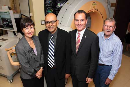
A $500,000 donation from BMO Financial Group has enabled The Neuro (Montreal Neurological Institute and Hospital) to install a vital surgical imaging tool, known as an O-arm Imaging System. This new technology allows neurosurgeons to perform advanced surgical procedures with maximum precision, enhance operating room efficiencies, and improve overall patient outcomes.
The O-arm combines intra-operative imaging with image-guided surgery (surgical navigation systems, analogous to a GPS system), providing real-time, high quality 3D images of a patient’s anatomy that are essential to surgeons during an operation. Each year, thousands of Canadians require surgical intervention to eliminate or control the symptoms of conditions such as epilepsy, brain tumours and Parkinson’s disease.
“For Parkinson’s patients, the difference in quality of life before and after surgery is enormous,” said Dr. Abbas Sadikot, neurosurgeon at The Neuro and Head of Functional Neurosurgery Service at the MUHC. Due to tremor, stiffness or slowness, Parkinson’s patients often have difficulty performing even the simplest tasks, such as picking up a glass of water or writing legibly on a piece of paper. But relief can come with the flip of a switch for some Parkinson’s sufferers. By surgically implanting devices called Deep Brain Stimulators (DBS) that electrically stimulate specific parts of the brain, symptoms of tremor, rigidity, stiffness, slowed movement and walking impairments can be dramatically relieved.
A crucial step towards a successful outcome is determining whether an implanted device has been correctly placed. “The O-arm is absolutely essential to these surgeries because it allows us to confirm that the DBS is in the correct location in the brain so patients don’t have to come back for a second surgical procedure for corrections and adjustments. Prior to acquiring the O-arm, we used post-operative MRI to check the results, and patients had to come back for revision surgeries,” said Dr. Sadikot.
Other advantages include: increased efficiency in the OR due to fast access to images – the O-arm takes just minutes to combine intra-operative CT with pre-op MRI – which also means increased safety due to minimized radiation exposure for surgical staff. The O-arm is used mainly in the treatment of Parkinson’s disease but it is also used with patients suffering from trigeminal neuralgia, cluster headaches and for some spinal surgeries.