By Fabienne Landry, MUHC Communications
Over the past 30 years, advancements in medical imaging have allowed for earlier diagnosis and improved patient outcomes. As a pediatric radiologist at the Montreal Children’s Hospital of the McGill University Health Centre (MCH-MUHC), Dr. Ricardo Faingold is well aware of this, but he also knows that there are many places in the world that lack the resources and know-how when it comes to radiology. Coming back from an outreach trip to Maputo, Mozambique, he told us how he uses his time and skills to help change that reality.
What was the main goal of the trip?
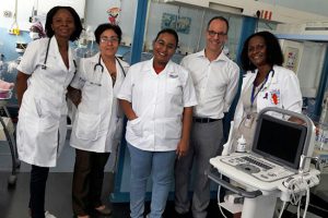
I went to Maputo Central Hospital to teach staff and residents in the Departments of Pediatrics, Neonatology and Radiology. It was an initiative of the World Federation of Pediatric Imaging (WFPI) in partnership with the University of California, Los Angeles (UCLA) Global Health Project. I spent a week there, teaching how to perform radiology exams, mostly ultrasounds, and giving various workshops and lectures on different topics.
Why ultrasound?
Ultrasound is a very useful diagnostic technology in pediatrics, first because it uses no radiation, and second, the equipment might be small and inexpensive equipment. With this you can do a lot for kids; for babies you can even perform head ultrasounds through the fontanel. Head ultrasound in premature infants is very important at the Neonatal intensive care unit (NICU) to diagnose and monitor many conditions that are life threatening.
What did you first do when you got there?
I arrived on Sunday night, November 27, and I was very well received by Dr. Chris Buck, a pediatrician in charge of UCLA Global Health, who has been working at the Maputo Central Hospital for three years now. The next morning, I met key people in the administration, organized the courses and lectures, and went to the NICU to show the care team how to perform ultrasound on premature babies. I was actually doing it and teaching it at the same time, and we examined all the babies who were there.
How did it go?
To my surprise, the ultrasound equipment they had in the NICU was still in its original boxes. The Chinese Government had donated it and since they had no biomedical engineer to assemble it, I was asked to help and immediately recruited one of the local residents for the task. After a few hours, the equipment was ready for use! Although challenging, this step was crucial because it allowed the NICU, which is located in a separate building from the Radiology and Pediatrics departments, to have its own equipment.
Overall, what were your main duties during your stay?
I gave a total of nine lectures, mainly to pediatricians and surgeons, about everything from x-rays to CT scans, MRIs and ultrasound. I taught them how to read the basics so that they could help the patients on a daily basis. I showed them how to identify pneumonias, complications of pneumonia, diseases that babies are born with, disease from the abdomen that may or may not require surgery, and more.
One of the mornings, I did ward rounds with Dr. Buck, residents and the team from general surgery. As you might expect, there were children with malnutrition facing very challenging social and economic realities. We diagnosed many conditions from tuberculosis to rare tumours, infections, abscesses, obstructions and twists in the bowel.
Did you have a chance to meet their local radiologists?
Radiologists form a very small group and none of them are pediatric experts. They are always extremely busy, so they did not have time to attend many lectures, but I was able to spend some time with them and pass some of my experience with pediatric radiology.
What was your main motivation to embark on this outreach trip?
In countries like Canada or the US, the practice of medicine today relies a lot on technology and medical imaging. For instance, here at the MUHC we are lucky to have many radiologists who can interpret the exams and perform procedures on a daily basis. If the surgeon calls us for something really urgent we can be available right away. They don’t have that flexibility everywhere. I believe we can improve health care in the world by making medical imaging more available.
I heard that you are from Brazil, so I guess speaking Portugese was also an asset on this trip?
Yes, of course. I was able to teach in their language and interact efficiently with patients and family.
Are you planning to work again with the WFPI?
I’d like to repeat the experience, for sure. In the meantime, I do what I can to help at a distance. Like many other volunteers who work with the WFPI, I sometimes read CT scans and X-rays that are sent to us from remote areas in Asia and Africa, where they have access to some imaging technologies but lack expertise to interpret the images. I do that on a regular basis, and I’m happy to help them take care of their patients. But in the end, the best thing we can do is teach them how to do it. We want to empower them, and this is why we need to keep doing these teaching trips and raising awareness within our own communities.
On a personal level, what does this type of work bring you?
When you teach people and they start using that new knowledge, you have that satisfaction of seeing the impact it has on patient care. We are not only helping those specific patients we saw during the trip. By transmitting our knowledge, we are in fact helping many more patients to come. I am proud to say that the NICU in Maputo Central Hospital can now perform daily basic head ultrasounds with its own equipment. We also created a personal connection with the health care professionals in that institution and that is very important.
